| Table of Contents | |
|
Case Report
| ||||||
| Severe tracheal stenosis after short-term endotracheal intubation: A case report | ||||||
| Tatjana Goranović1, Zoka Milan2, Irena Pirkl3, Višnja Nesek Adam4 | ||||||
|
1MD, PhD, Senior Teaching Assistant, Consultant Anesthetist and Intensivist, Department of Anesthesiology, Reanimatology and Intensive Care Medicine, University Department for Tumors, Sestre milosrdnice University Hospital Centre, Zagreb, Croatia; Senior Teaching Assistant, Faculty of Medicine, University of Osijek, Croatia.
2MD, PhD, Honorary Senior Lecturer, Visiting Professor, Consultant Anesthetist and Intensivist, Honorary Senior Lecturer, Visiting Professor, Anesthetic Department, King's College Hospital, London, United Kingdom. 3MD, Consultant Otorhinolaryngologist, Department of Otorhinolaryngology and Head and Neck Surgery, Sveti Duh University Hospital, Zagreb, Croatia. 4MD, PhD, Assistant Professor, Consultant Anesthetist and Intensivist, Head of University Department of Anesthesiology, Reanimatology and Intensive Care Medicine, Sveti Duh University Hospital, Zagreb, Croatia; Assistant Professor, Faculty of Medicine, University of Osijek, Croatia. | ||||||
| ||||||
|
[HTML Abstract]
[PDF Full Text]
[Print This Article]
[Similar article in Pumed] [Similar article in Google Scholar] |
| How to cite this article |
| Goranovic T, Milan Z, Pirkl I, Nesek Adam V. Severe tracheal stenosis after short-term endotracheal intubation: A case report. Edorium J Anesth 2016;2:10–13. |
|
Abstract
|
|
Introduction:
Prolonged endotracheal intubation is a risk factor for the development of tracheal stenosis. The incidence of stenosis is very low if intubation lasts less than a week and patients may be asymptomatic for a long time.
Case Report: We present a case of an 86-year-old female who developed severe tracheal stenosis after short-term endotracheal intubation, with her first hospital admission for stridor only two weeks after the intubation. One month after the intubation computed tomography (CT) scan revealed an 18-mm long tracheal stenosis at the level of the thyroid gland, 1 cm below the glottis, with 3 mm of free tracheal lumen at the narrowest part. During CT scan, the patient rapidly became dyspnoeic, cyanotic, and agitated. An urgent tracheostomy was performed under local anesthesia with the patient in a semi-sitting position. When ventilation through this tube was possible, the patient was anesthetised, repositioned in the supine position and a permanent tracheostomy was performed. Conclusion: An atypical medical history delayed the diagnosis and treatment, which would have been different if the appropriate diagnosis had been made earlier. Presentation with symptoms of airway obstruction and a history of mechanical intubation, no matter how short and recent, requires detailed history taking, careful examination and diagnostic approach to make the diagnosis of tracheal stenosis in a timely manner and treat it adequately. | |
|
Keywords:
Endotracheal intubation, Mechanical ventilation, Stridor, Tracheal stenosis, Treatment
| |
|
Introduction
| ||||||
|
Although endotracheal intubation is a common procedure in intensive therapy units, it still qualifies as an invasive procedure. Prolonged endotracheal intubation is a risk factor for the development of tracheal stenosis [1] [2][3]. The most likely mechanism of tracheal stenosis is an injury of tracheal mucosa, with secondary scar healing that consequently causes the development of some degree of tracheal stenosis [4]. Depending on the degree of tracheal stenosis, patients may be asymptomatic for a long time or symptoms can occur within several months [5]. The incidence of stenosis is very low if intubation lasts less than a week [4]. We experienced a patient who developed severe tracheal stenosis after short-term endotracheal intubation, with her first presentation only two weeks after the intubation. An atypical medical history delayed the diagnosis and treatment, which would have been different if the appropriate diagnosis had been made earlier. | ||||||
|
Case Report
| ||||||
|
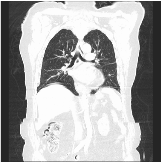
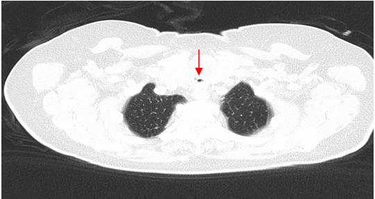
An 86-year-old woman presented to the accident and emergency department (A&E) with stridor. She was not able to talk in clear sentences. In addition, lung auscultation revealed weak breathing and prolonged expiratory phase. Initial arterial blood gases analysis on air was as follows: pH 7.302, PaCO2 6.75 kPa, PaO2 12.69 kPa, base excess -1.3, standard bicarbonate 23.3 mmol/L, actual bicarbonate 25.3 mmol/L, total bicarbonate 26.8 mmol/L, SaO2 0.96. This was her third presentation to A&E with symptoms of airway obstruction within four weeks. On the two previous admissions, she had been treated with bronchodilators and discharged home after brief observation. The patient had a complex past medical history, including a high body mass index (BMI), a history of severe hypertension, atrial fibrillation, chronic bronchitis and pulmonary hypertension. One month earlier, she had an episode of pulmonary edema and cardiac arrest and was briefly intubated with 7.5 mm polyethylene endotracheal tube (ETT) and ventilated 53 hours. Seven days after the cardiac arrest, she recovered completely and was stable enough to be discharged home. Since this was her third presentation with the same symptoms, an ear nose throat (ENT) consultation was requested and computed tomography (CT) ordered (Figure 1). The CT scan revealed an 18-mm-long tracheal stenosis at the level of the thyroid gland, 1 cm below the glottis, with 3 mm of free tracheal lumen at the narrowest part (Figure 2). During CT scan, the patient rapidly became dyspnoeic, cyanotic, and agitated. The anesthesiologist was called, while ENT consultant indicated tracheotomy. The patient was transported to the operating theatre without any other further testing. An urgent tracheostomy with a 3.5 mm ETT was performed under local anesthesia with the patient in a semi-sitting position. When ventilation through this tube was possible, the patient was anesthetized, repositioned in the supine position and a permanent tracheostomy was performed. The patient was fully awake at the end of surgery and transported to intensive care unit for 24 hours monitoring. The external thoracic surgeon was consulted for possible further tracheal reconstruction. However, because of the anatomical position of the stenosis and the patient's age and comorbidities, further tracheal surgical reconstruction with end- to- end anastomosis was contraindicated and tracheal stoma was left as permanent and the patient discharged home. | ||||||
| ||||||
| ||||||
|
Discussion
| ||||||
|
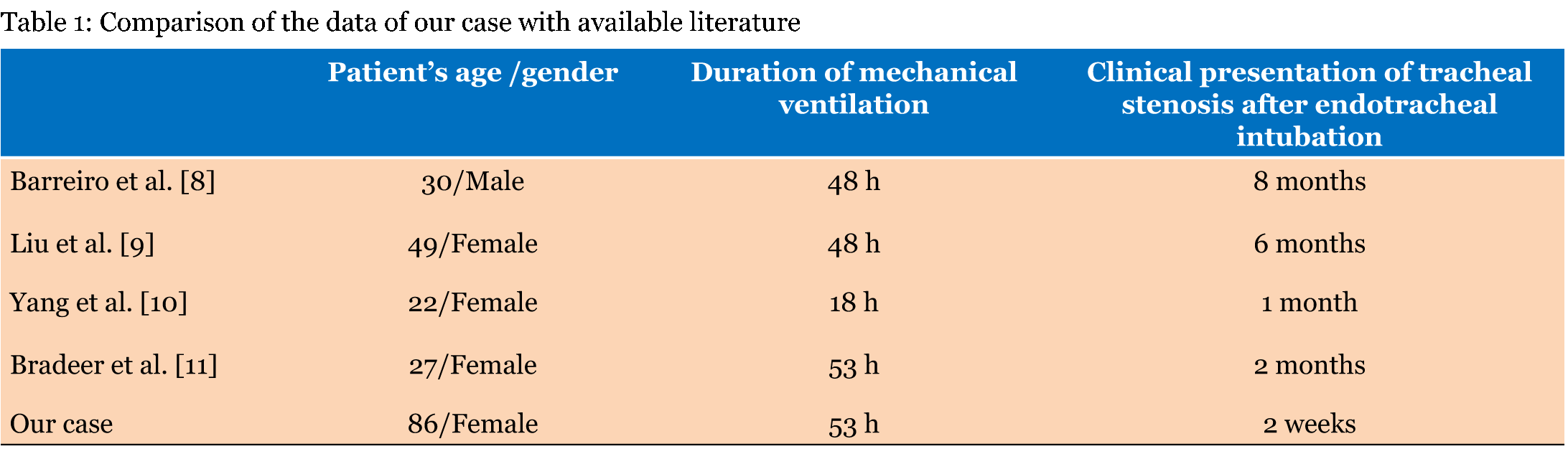
We present an elderly patient who presented to A&E with stridor three times before a diagnosis of severe tracheal stenosis was made. During diagnostic CT scan, the patient became hypoxic and an urgent tracheostomy was performed, followed by a permanent tracheostomy. The high (sub-glottic), 18-mm-long, severe (3-mm-diameter) stenosis was probably caused by a 53-hour period of mechanical ventilation. Her first symptoms occurred two weeks after this. This case is noteworthy because of the early symptom onset and severe stenosis after a short period of mechanical ventilation. The patient was not able at the time of admission to provide a full medical history and she was discharged twice from A&E without a proper diagnosis. When she was finally diagnosed, she became respiratory critical and required an urgent tracheostomy. Reported aetiological factors for tracheal stenosis include the duration of intubation, cuff pressure acting on the tracheal mucosa, traumatic intubation, hypotension at the time of intubation, obesity, diabetes mellitus, advanced age, excessive corticosteroid steroid use, female and oestrogen effect, comorbidities [severe respiratory failure, severe reflux disease, autoimmune diseases (Wegener's granulomatosis, sarcoidosis and others)], obstructive sleep apnoea, and smoking history [1] [6] [7]. The most common profile of a patient with tracheal stenosis in one series of 31 patients was a female (75%), obese (66%) patient with a history of diabetes mellitus (35.4%), hypertension (51.6%), and cardiovascular disease (45.1%), who was a current smoker (38.7%) [1]. Our patient had five risk factors: urgent intubation, hypotension at the time of intubation, advanced age, obesity and female. Unfortunately in our case, we could not check the details about intubation since this had happened in another hospital. A high cuff pressure acting on the tracheal mucosa (>30 mmHg) may cause regional ischemia within 15 minutes that can lead to ulceration, chondritis, and eventually fibrosis in 3–6 weeks [8]. Despite no clear note in medical documentation about measuring the cuff pressure, we think that it is less likely that high cuff pressure would attribute to the trauma in our case. The ET tubes in current use are high-volume, low-cuff pressure tubes, which should lower the risk of tracheal stenosis. But we may highly assume that intubation was rather traumatic as this was the case of resuscitation. Patient's cardiovascular status and low regional perfusion could attribute to regional ischemic damage of tracheal mucosa at the time of intubation because she was in cardiac arrest. Our patient's gender (female) might influence fibrosis formation, but this attributes to female sex hormones that were less relevant in this case as the patient was menopaused for decades. There are only a few reports on tracheal stenosis developing after intubation in less than 6 days (Table 1) [8][9][10][11]. The number of reported cases has been too small so far to make define clear cut off regarding hours of intubation that cause the local tissue damage or timing of critical fibrosis development. However, our case of early development tracheal stenosis is specific for the earliest clinical presentation of tracheal stenosis (only two weeks). Our patient's age contraindicated eventual tracheal surgical reconstruction, which is the standard of care of tracheal stenosis, with success in up to 96% of cases [3]. However, there are less invasive treatment modalities, such as laser excision [12] , cryotherapy [9], balloon dilatation and stenting[2] [13], and topical mitomycin C [14]. We made a definitive diagnosis of post-intubation tracheal stenosis at a critical moment, although the patient had milder symptoms on her two previous visits to A&E. If the stenosis had been suspected earlier, an optional less invasive treatment of the tracheal stenosis (e.g. laser excision or topical mitomycin C) that was more appropriate for the patient's age might have been possible. | ||||||
| ||||||
|
| ||||||
|
Conclusion
| ||||||
|
Presentation with symptoms of airway obstruction and a history of mechanical intubation, no matter how short and recent, requires a multidisciplinary approach in order to diagnose tracheal stenosis in a timely manner and treat it adequately. Geriatric patients with a history of mechanical ventilation after resuscitation are more likely to develop post-intubation tracheal stenosis in a relatively short time after the critical incident. | ||||||
|
References
| ||||||
| ||||||
|
[HTML Abstract]
[PDF Full Text]
|
|
Author Contributions
Tatjana Goranović – Substantial contributions to conception and design, Acquisition of data, Analysis and interpretation of data, Drafting the article, Revising it critically for important intellectual content, Final approval of the version to be published Zoka Milan – Substantial contributions to conception and design, Analysis and interpretation of data, Drafting the article, Revising it critically for important intellectual content, Final approval of the version to be published Irena Pirkl – Acquisition of data, Analysis and interpretation of data, Drafting the article, Revising it critically for important intellectual content, Final approval of the version to be published Višnja Nesek Adam – Acquisition of data, Analysis and interpretation of data, Drafting the article, Revising it critically for important intellectual content, Final approval of the version to be published |
|
Guarantor of submission
The corresponding author is the guarantor of submission. |
|
Source of support
None |
|
Conflict of interest
Authors declare no conflict of interest. |
|
Copyright
© 2016 Tatjana Goranović et al. This article is distributed under the terms of Creative Commons Attribution License which permits unrestricted use, distribution and reproduction in any medium provided the original author(s) and original publisher are properly credited. Please see the copyright policy on the journal website for more information. |
|
|